baby chest x ray holder
Chest X-ray or chest radiograph is the most commonly performed diagnostic imaging test. Chest x-ray is the most commonly used imaging exam for evaluating the chest.
Paediatric Chest Immobilisation Devices Wikiradiography
The device is the size of a small camera.

. They may take 2-3 days. X-rays are used throughout the body. It turns out this photo originally posted on Reddit of a baby squished into a tube like a lil baby deposit is actually of him getting a tiny.
It is often the first type of imaging used to identify sources of pain evaluate traumatic injuries and locate a foreign body. Your child will need to hold very still while the X-ray is taken. Your child may be asked to hold his or her breath for a few seconds.
Your child stands or lies down with his or her chest against an X-ray plate for the pictures. During the examination an X-ray machine sends a beam of radiation through the chest and an image is recorded on special film or a computer. Adjustable seat platform can be rotated horizontally to any degree and position to facilitate oblique and lateral views.
4 x 4s b. If the childs like less than a month old has a high fever a white blood cell. A pediatric chest X-ray is a non-invasive procedure that provides a picture of a childs heart lungs and bones in the chest.
Soft stretchy Velcro straps are used to secure the arms in the desired pose. C-Spine Chest Abdominal Magnified Airway Soft Tissue Neck. Once you get the chest x-ray report of your child you may start wondering about some guidance from the radiologist.
The Pigg O Stat is a fast sure and simple method for immobilizing and positioning children from infants to about 3 years of age. Punger and she wanted us to get a chest xray this afternoon then a visit to her office. While not completely comfortable for the kid it doesnt hurt.
Its called a Pigg-O-Stat. A chest X-ray is a safe and painless test that uses a small amount of radiation to take a picture of a persons chest. Ad Browse discover thousands of brands.
It has a seat like a bicycle saddle that the baby straddles. Getting the chest x-ray results. Lateral chest and abdomen views.
Like other radiography methods taking images with X-ray involves exposing a part of the body to a small ionizing radiation dose 2. Cassettes must be entirely stationary to acquire the most detailed image so it would not be wise to allow patients or personnel to hold them in place. By the way this is mainly for trauma.
CHEST TUBE SIZE ETT x 4 If the same six yo patient needs a chest tube your chest tube size is 55 x 4 22 Fr. This noninvasive method produces images of the heart lungs airways blood vessels and the bones of the chest and spine 1. It shows the location size and shape of the heart lungs and blood vessels.
A chest X-ray can be useful in the diagnosis of pneumonia tumors collapsed lung. Small of transparent Plexiglas and an adjustable. ETT x 3 If your calculated tube size is 55 your depth should be 55 x 3 165 cm.
It has wires with silver dollar-sized electrodes that attach to your skin. When the chest radiograph also includes the abdomen look out for the umbilical clip. It should ideally be performed in the ICU using a portable X-ray equipment.
So 165 cm mark should be seen at the level of central incisors. It might look scary but it. They are an excellent tool for reducing patient exposure because they reduce motionblur thereby reducing the need for repeat exposures.
Unfortunately Nati is still sick with a nasty cough and congestion. A Holter monitor is a battery-operated portable device that measures and records your hearts activity ECG continuously for 24 to 48 hours or longer depending on the type of monitoring used. These are plastic clips used to clamp the umbilicus before it is cut at birth.
We made it to the radiologist office where my sweet bundle of joy transformed into a seemingly 8-legged octopus squirming madly. This image includes organs and structures such as the heart lungs large blood. 2 x 2s c.
Your child will be given a gown to wear during the test. X-ray exams are used to help diagnose a wide variety of injuries and illnesses in children. Instead use a mobile or table top cassette holder so the patient may relax and the employee can stand at a safe distance.
We spoke with Dr. Still in a minority of cases that are more complex an X-ray may be helpful. The umbilical stump remains in situ for approximately 1-2 weeks and.
In most cases an anteroposterior view of the chest would provide sufficient diagnostic information. Its generally used for chest radiographs. The pigg-o-stat includes form-fitting body and arm supports one pair each - large.
The chest X-ray is the most frequently ordered radiological investigation in NICUs. Read customer reviews find best sellers. In case of any emergency the chest x-ray are handed over on the same day else.
Pediatric X-Ray Positioning for Infants. The chin strap is also used to support the head and neck. Its use is to immobilize the child so that they dont wiggle around.
The infant saddle seat is used for infants through about 8 month olds. Curved Kelly clamp f. X-ray CR cassette holders are necessary to position the cassettes in place sturdily.
But the real and accurate analysis is done by the child doctor. I use one every time I do a chest X-ray on a baby. Curved mosquito hemostat e.
It can also show a doctor if there is fluid in the lungs. Needle holder 6 Sterile thoracotomy tube 12F for infants greater than 1500 grams and 10F for infants less than 1500 grams or Sterile percutaneous safety pneumothorax system or Sterile 85 French pigtail catheter 7.
![]()
Pediatric Cxrs A Normal Cxr Showing Clear Lungs With No Abnormal Download Scientific Diagram
Radiology In Ped Emerg Med Vol 1 Case 8

Chest X Ray Desk Organizer Zazzle Com Desk Organization White Ceramic Tiles Tile Design
Paediatric Chest Immobilisation Devices Wikiradiography

Chest X Ray In The 5 Th Month Of Att Partial Resolution Of The Download Scientific Diagram

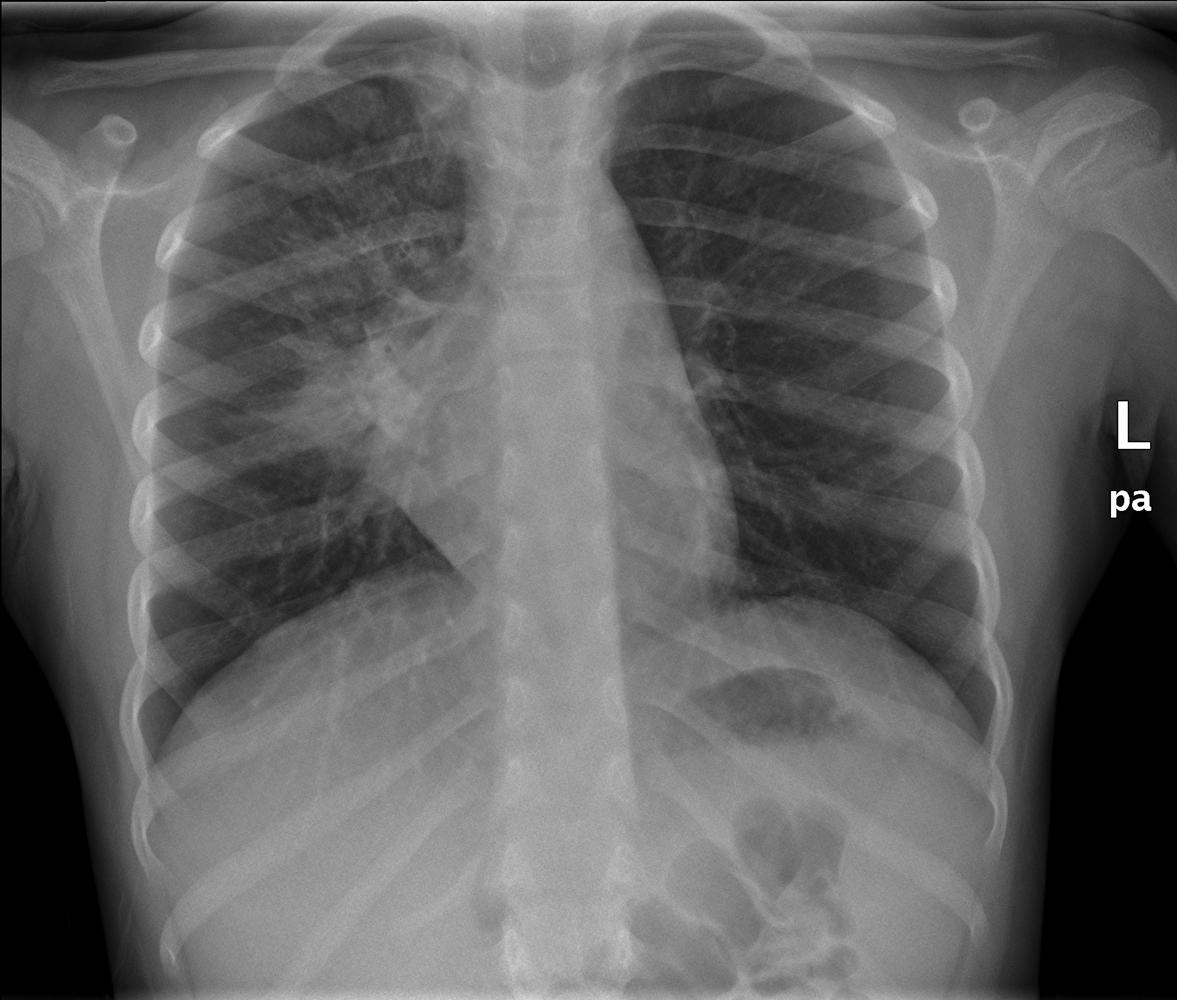
Chest Radiograph Paediatric Radiology Reference Article Radiopaedia Org

Chest X Ray Confirming Adequate Placement Of Esophageal Cooling Device Download Scientific Diagram

Normal Chest X Ray Throw Blanket Cafepress Lunges Fun Workouts X Ray

Hangman S Fracture Xray Xr Cervical Spine Vertebrae Clay Shoveler S Fx Forward Head Posture Posture Fix Sternocleidomastoid Muscle
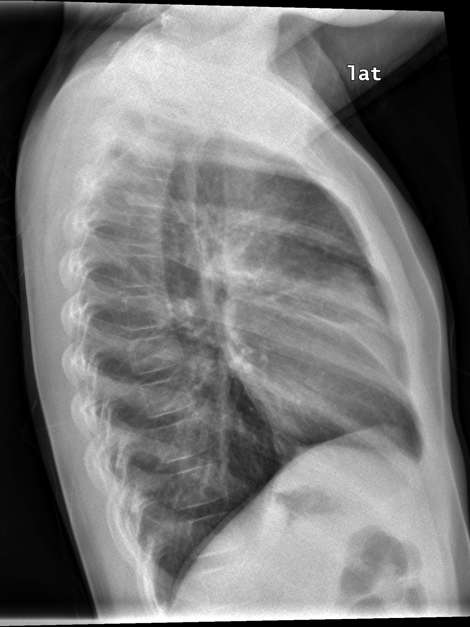
Paediatric Chest Immobilisation Devices Wikiradiography
Enlargement Of The Cardiac Silhouette On Chest X Ray Lecturio
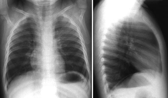
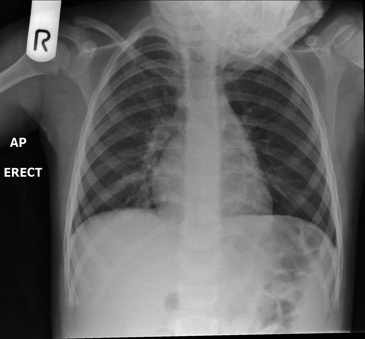
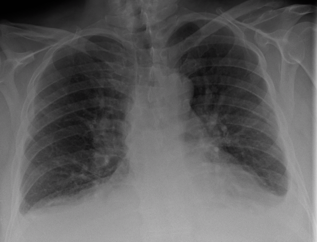
Pediatric Chest Ap Erect View Radiology Reference Article Radiopaedia Org

This Is What It Looks Like When A Baby Gets An X Ray X Ray Radiology Humor Cute Babies

Pediatric Chest Supine View Radiology Reference Article Radiopaedia Org

Neonate Chest Supine View Radiology Reference Article Radiopaedia Org
![]()
Osteopoikilosis Spotted Bone Disease Radiology Imaging Nuclear Medicine Technology Bone Diseases

Chest Xray Of A Child With A Cough Shows Pneumonia Radiologist Radiology Diagnostic Imaging Medical Imaging X Ray

Chest X Ray Desk Organizer Zazzle Com Desk Organization White Ceramic Tiles Tile Design

Chest Xray In A Child With Cough Shows Round Pneumonia Radiologist Radiology Radiology X Ray Radiologist